3. Applications
It is of course impossible to give a complete overview of the large number of applications that have used optically enhanced magnetic resonance techniques in various systems. The selection that is presented here tries to demonstrate the wide range of syst ems for which optical methods can be used, but in addition also the various ways in which optical radiation can enhance magnetic resonance spectroscopy.
3.1. Dielectric Solids
Optical pumping and optical detection of magnetic resonance transitions were first demonstrated in atomic vapours [Brossel, Kastler, 1952, Kastler, 1967] but soon afterwards also in ionic solids [Geschwind, Collins, 1959, Wieder, 1959]. Ruby (Cr 3+:Al 2O 3) proved an ideal test material, as it has relatively narrow optical absorp tion lines and allows different methods of optical pumping. Most experiments reported so far were performed with inorganic ionic solids, in particular rare earth and transition metal compounds. The experimental techniques used for these studies include purely optical methods like photon echo modulation [Chen, Chiang, 1980, Szabo, 1986] and coherent Raman beats [Brewer and Hahn, 1973] as well as double resonance methods like Raman heterodyne spectroscopy [Wong, Kintzer, 1983].
We start the discussion with an example for a purely optical method that combines several attractive features: while its sensitivity is that of an optical method, its resolu tion is not limited by laser jitter or optical dephasing, but only by the natural linewidth. Furthermore, it yields magnetic resonance spectra from an elec tronically excited as well as from the ground state. The material system that we consider here consists of the rare earth ion 141Pr 3+ with a nuclear spin I = 5/2. The ions are substituted into the host material of YAlO 3, where they occupy the Y sites with a doping concentration of 0.1 %. In zero magnetic field, the nuclear spin states of 141Pr are split by the interaction between the nuclear quadrupole moment and the electric field g radient (EFG) tensor; the interaction with the quenched electronic spin enhances the quadrupole coupling as well as the nuclear Zeeman interaction.
The experimental scheme uses Raman processes for exciting the nuclear spin
transitions as well as for observing the spin precession. A laser pulse that
couples to two optical transitions sharing a common energy level excites a cohe
rence in the third transition of the three level system, as explained above. The
excitation pulse should not be longer than the spontaneous lifetime of the
electronic state that is to be investigated. For the 1D 2 state of Pr 3+, this
limits the duration of the laser pulses to t P < 100 msec. After the pulse,
the coherence is allowed to precess freely and a second, weaker laser beam is
used to observe the precession, again using a Ra man process. The condition for
efficient excitation of the sublevel coherence is that the difference between
the two frequency components of the exciting laser pulse is close to the
sublevel transition frequency [ Blasberg and Suter, 1994] . For the observation
process, the laser frequency must be close to one of the two optical transition
frequencies. The signal which is observed after the optical pulse can be Fourier
transformed to reco ver the usual NMR / NQR spectrum. Figure 3.1 shows two examples of spectra that can be obtained with this method.
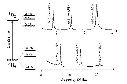
Figure 3.1. Example of NQR spectra obtained by purely optical excitation and detection of nuclear spin coherence. Both spectra were combined from three partial spectra which were recorded with different modulation frequencies to reduce distortion due to finite excitation range.
They were obtained from the electronic ground state and an electronically excited state of Pr 3+:YAlO 3. For both states, not only the magnetic dipole-al lowed transitions (+/-1/2 ' +/-3/2, +/-3/2 ' +/-5/2) are visible, but also the double quantum transition (+/-1/2 ' +/-5/2), indicating that the selection rules of conven tional magnetic resonance do not apply to Raman-detected magnetic resonance. The quadrupole interaction strength differs significantly for excited and ground state (0.9 MHz vs. 7 MHz). Like with magnetically excited spectra, the band width of the excitation process is limited. In this case, the transition strength is quite low. With a laser intensity of 20 mW, it was then possible to excite subspectra with a width of approximately 1 MHz. To reduce the distortions associ ated with the excitation of considerably wider spectra, we combined therefore three subspectra containing one resonance line each. The width of the resonance lines is determined by spin diffusion of the dipole-dipole interaction with the neigh bouring Al nuclei.
Excitation of magnetic resonance transitions with radio frequency irradiation relies on the conversion of longitudinal into transverse magnetisation; an experiment can therefore start only after thermal relaxation has established popula tion differences by coupling to the lattice. At low temperatures, this process can be very slow in rigid solids. Optical excitation, in contrast, converts population differences between different electronic states into transverse magnetisa tion. Spontaneous emission establishes these population differences on a very short time scale (ns to ms), independent of temperature. As a result, purely optical experiments do not require long relaxation delays and experi ments can be repeated very quickly.
Raman experiments do not have to use only optical techniques. Double resonance techniques that include optical as well as rf irradiation have been used successfully. The rf field is then used to establish a coherence in a magnetic reso nance transition, which is detected with the same Raman process that we discussed above. In such an experiment, the laser beam serves a threefold pur pose: it establishes a population difference which is converted into transverse magnetisation by the rf field. The same [Wong, Kintzer, 1983] or a second [ Blasberg and Suter, 1993] laser beam converts the precessing magnetisation partia lly into optical polarization by a coherent Raman process, and as a third function, the laser beam serves as the local oscillator for the detection of the Raman field. This technique was first used to observe NMR transitions in rare earth compounds [ Mlynek, Wong, 1983, Wong, Kintzer, 1983] and later also to ESR [Holliday, He, 1990] and ENDOR [Manson, He, 1990] [Manson, Fisk, 1992].
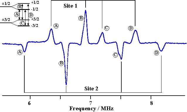
Figure 3.2. Raman heterodyne spectrum of Pr 3+ :YAlO 3 in a weak magnetic field.
Figure 3.2. shows the Raman heterodyne signal, again from Pr 3+:YAlO 3,
as a function of the fre quency of the rf field.
The spectrum shown here was measured in a weak magnetic field of 7 mT, so that the transitions between the +1/2 and +3/2 states are no longer degenerate with the transition from the -1/2 to the -3/2 state.
The quadrupole coupling and the (enhanced) Zeeman interaction are strongly anisotropic.
For a general orientation of the magnetic field with respect to the quadrupole coupling,
the usual dipole selection rules of high-field NMR are not applicable, and also the transitions
between the +1/2 and -3/2 and between the -1/2 and +3/2 states are partially allowed.
A given spin has therefore 4 possible transitions between these states, as indicated in the inset.
In addition, the crystal has 2 non-equivalent sites, resulting in a total of 8 resonance lines.
Again, the width of these resonance lines is independent of the laser linewidth and only due to properties of the crystal.
In these experiments, the laser beam interacts directly with the atom whose nuclear spin transitions are measured. In other cases, spin-spin couplings exchange polarizations with neighbouring spins. It is then possible to optically pola rize those spins and detect their NMR transitions optically [Szabo, Muramoto, 1990, Erickson, 1993]. In other cases, the interaction between two neighbouring atoms manifests itself more strongly in the optical transition. The position of the opt ical resonance within the inhomogeneously broadened resonance line could then be used to assign the magnetic resonance spectrum to a pair of neighbouring spins [Lukac and Hahn, 1989] . While most experiments of this type were performed in ionic materials, colour-centres in diamond have also been studied [Manson, He, 1990].
3.2. Semiconductors
Semiconductors have become a very active area for applications of optically enhanced magnetic resonance. The first demonstrations of the technique were performed in GaAs [Paget, 1981] . For the optical excitation, one usually chooses a photon energy at the exciton resonance or just above the band gap, thereby creating holes at the top of the valence band and electrons at the bottom of the conduction band. Under these conditi ons, the system behaves to some degree like free atoms. Figure 3.3. shows the relevant band structure for the technically important case of a III-V semiconductor with tetrahedral symmetry.

Figure 3.3. Optical Pumping in semiconductors: level scheme for a III-V compound with tetrahedral symmetry.
In these compounds, the valence band is composed of p-type orbitals (L=1). Spin-orbit coupling splits the band into a J=1/2 and a J=3/2 subband. The conduc tion band consists of s-type orbitals and the total electronic angular momentum of excited electrons is therefore J=1/2. Absorption of a circularly polarized photon by an electron in the J=1/2 valence band creates then a hole and a con duction band electron, both with J z = +1/2. In the valence band as well as in the conduction band the electron spin system becomes then polarized to some degree that depends on the thermal isation times of hole and electron. Like in free atoms, the spin polarization reflects itself in the polarization of the luminescence.
The hyperfine interaction between electrons and nuclear spins can transfer spin polarization from the electrons to nuclei. If the nuclear spin reservoir is perturbed by resonant rf irradiation, the electron spin polarization and there fore the polarization of the luminescence are also affected. By observing the luminescence and scanning the radio frequency in a constant magnetic field, it is thus possible to record magnetic resonance spectra [Paget, 1981, Krapf, Denninger, 1991].

Figure 3.4. Example of optically detected NMR spectrum of GaAs. The signal was re corded by scanning the radio frequency and observing the luminescence in a constant magnetic field of 0.17 T.
Figure 3.4. shows an example of such a spectrum from GaAs, reproduced from ref. [Krapf, Denninger, 1991]. Two different isotopes of Ga are clearly visible, as well as the As reso nance. The positions of the resonance lines depend on the polarization of the electron spins, as shown in the lower left part of the spectrum. Such experiments may well become an interesting addition to the ana lytical tools of the semiconductor industry. Most experiments performed up to now were directed on GaAs, but Si, Ge [Glaser, Trombetta, 1990], and InSb [Hofmann, Pascher, 1993] have also been investigated.
Apart from the polarization and intensity of the light, NMR transitions can be observed indirectly also through changes in the ESR resonance position [Hofmann, Denninger, 1993]: the cou pling between the electronic and nuclear spins shifts the ESR transition frequency. By applying pulsed or cw radio frequency fields to the nuclear spins, it is possible to change their polarization and thereby the ESR frequency. If the number o f spins is large enough, it is even possible to observe the NMR spectrum directly in a conventional spectrometer modified for optical pumping [Barrett, Tycko, 1994].
The high sensitivity of optical techniques is a very attractive feature for studying the thin films used in integrated circuits. The optical techniques could become even more attractive for studying quantum confined structures [Uenoyama and Sham , 1990]. When the conduction electrons are confined to small regions, like quantum wells, quantum wires or quantum dots, the optical absorption spectra change considerably. It is then possible to excite selectively on ly those areas, where the electrons are confined and thereby get spectroscopic information specifically from those areas. Such experiments require therefore not only high sensitivit y for measuring signals from small parts of the sample, but also good selectivity to extract this signal from the large background of the whole rest of the sample.
3.3. Surfaces
Quasi-twodimensional systems have always proved difficult to investigate by NMR, since the number of spins in these systems is quite small [Duncan and Dybowski, 1981, Slichter, 1986] . Increasing the spin polarization by optical pumping has significantly improved the sensitivity of this type of experiments. In particular the transfer of spin polarization from optically pumped alkali atoms to Xe nuclear spins [Happer, Miron, 1984] has allowed to study the effect of surfaces on the magnetic resonance spectrum [Wu, Happer, 1990, Butscher, Waeckerle, 1994]. In the fast exchange regime, the distribution of the spins between surface adsorbate and va pour phase determines the averaged magnetic resonance spectrum. For oriented surfaces, where most spins are in the vapour phase, the splittings and shifts are then in the mHz range, requiring highly sensitive detection meth ods. Other work has therefore concentrated on systems with large surface to volume ratio like Zeolites [Chmelka, Raftery, 1991], where most of the atoms are close to the interface.
For experiments with oriented surfaces, the use of light for optical pumping as well as for detection brings, apart from the sensitivity advantages, also the possibility to select signal contributions that originate from atoms that are close to the surface. For this purpose, changes in the penetra tion depth of light with wavelength [Lepine, 1972] has been used, but more frequently, the selection is achieved by reflecting a laser beam from the interface being investigated.
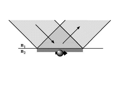
Figure 3.5. Principle of optical detection of magnetic resonance close to a surface. n 1,2 are the refractive indexes on both sides of the interface.
The reflection coefficient for the laser beam depends on the refractive index es on both sides of the interface and is therefore affected by atoms close to the interface that are resonant with the laser light. The changes in the reflection coefficient, which can be measured through changes of the amplitude and polariza tion of the reflected beam contain therefore information on the atoms close to the interface. The combination of optical pumping with this type of optical detection provides sufficient sensitivity that it is no longer necessary to use sam ples with high surface to volume ratios and allows therefore studies on oriented surfaces.
One method that relies on such a technique was used to study nuclear quadrupole resonances of Pr 3+ in LaF 3 [Lukac and Hahn, 1988] . In this case, the beam was reflected from an optically dense material. The reverse is also possible: if the laser beam undergoes total internal reflection at an interface to an optical ly less dense medium, an evanescent wave penetrates into the thinner medium by a distance of the order of the optical wavelength. Atoms in this evanescent wave can thus modify the reflected laser beam by absorbing li ght from it and by their effect on the refractive index and thereby on the reflection coefficient [Suter, Aebersold, 1991].
3.4. Molecules
Conventional magnetic resonance has been applied most successfully to molecular systems. These systems can be investigated also with optical methods. Aroma tic organic molecules are particularly suitable because their chromophores make optical spectroscopy particularly attractive.
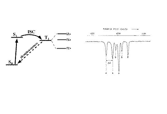
Figure 3.6. Principle of optically detected magnetic resonance in excited triplett states.
Experiments with these systems typically use optical excitation from the ground state to an excited singlett state with a pulsed UV laser. From the ex cited singlett state, intersystem crossing (isc) can populate a nearby triplet state. The intersystem crossing populates the different levels of the triplet state unequally. In addition, the triplet substates have in gene ral different lifetimes. Polarisation and intensity of the phosphorescence depend on the population of the individual states and allow an assessment of the population differences. By applying rf fields to the system it is possible to in duce transitions between the different triplet states; these transitions can be observed in the phosphorescence. Similar experiments have been performed on many other systems. The high sensitivity of the method was demonstrated very striking ly by to groups who measured magnetic resonance of individual pentacene molecules in p-terphenyl hosts [Koehler, Disselhorst, 1993, Wrachtrup, Borczyskowski, 1993]. Work is now in progress to extend these experiments to NMR transitions.
References
- S.E. Barrett, R. Tycko, L.N. Pfeiffer, and K.W. West, 'Directly-detected nuclear magnetic resonance of optically-pumped GaAs quantum wells', Phys. Rev. Lett. 72, 1369-1371 (1994).
-
T. Blasberg and D. Suter, 'Determination of the absolute sign of nuclear quadrupole interactions by laser - radio frequency double resonance experiments', Phys. Rev. B 48, 9524-9527 (1993).
-
T. Blasberg and D. Suter, 'Excitation of coherent Raman beats in rare earth solids with a bichromatic laser field', Optics Commun. 109, 133-138 (1994).
-
R.G. Brewer and E.L. Hahn, 'Coherent Raman beats', Phys. Rev. A 8, 464-472 (1973).
-
J. Brossel, A. Kastler, and J. Winter, 'Generation optique d'une inégalité de population entre les sous-niveaux Zeeman de l'état fondamental des atomes', J. Phys. Radium 13, 668-668 (1952).
-
R. Butscher, G. Waeckerle, and M. Mehring, 'Nuclear quadrupole interaction of highly polarized gas phase 131Xe with a glass surface', J. Chem. Phys. 100, 6923-6933 (1994).
-
Y.C. Chen, K. Chiang, and S.R. Hartmann, 'Spectroscopic and relaxation character of the 3 P 0 - 3 H 4 transition in LaF 3 :Pr 3+ measured by photon echoes', Phys. Rev. B 21, 40-47 (1980).
-
B.F. Chmelka, D. Raftery, A.V. McCormick, L.C. DeMenorval, R.D. Levine, and A. Pines, 'Measurement of Xenon distribution statistics in Na-A Zeolite cavities', Phys. Rev. Lett. 66, 580 (1991).
-
T.M. Duncan and C. Dybowski, 'Chemisorption and surfaces studied by nuclear magnetic resonance spectroscopy', Surf. Science Reports 1, 157 (1981).
-
L.E. Erickson, 'Nuclear-quadrupole-resonance measurement of the 27 Al frozen core in YAlO 3 :Pr 3+ ', Phys. Rev. B 47, 8734-8738 (1993).
-
S. Geschwind, R.J. Collins, and A.L. Schawlow, 'Optical detection of paramagnetic resonance in an excited state of C r3 + in A l 2 O3 ', Phys. Rev. Lett. 3, 545-548 (1959).
-
E. Glaser, et al., 'Detection of magnetic resonance on photoluminescence from a Si/SiGe strained-layer superlattice', Phys. Rev. Lett. 65, 1247-1250 (1990).
-
W. Happer, E. Miron, S. Schaefer, D. Schreiber, W.A.v. Wijngaarden, and X. Zeng, 'Polarization of the nuclear spins of noble-gas atoms by spin exchange with optically pumped alkali-metal atoms', Phys. Rev. A 29, 3092-3110 (1984).
-
W. Hofmann, G. Denninger, and H. Pascher, 'Investigation of the nuclear-spin polarization in InSb via spin-flip Raman gain spectroscopy', Phys. Rev. B 48, 17035-17042 (1993).
-
W. Hofmann, H. Pascher, and G. Denninger, 'Nuclear spin polarization in InSb detected by spin-flip Raman gain spectroscopy', Semicond. Sci. Technol. 8, S309-S312 (1993).
-
K. Holliday, X.F. He, P.T.H. Fisk, and N.B. Manson, 'Raman heterodyne detection of electron paramagnetic resonance', Optics Lett. 15, 983-985 (1990).
-
A. Kastler, 'Optical methods for studying Hertzian resonances', Science 158, 214-221 (1967).
-
J. Koehler, J.A.J.M. Disselhorst, M.C.J.M. Donckers, E.J.J. Groenen, J. Schmidt, and W.E. Moerner, 'Magnetic resonance detection of a single molecular spin', Nature 363, 242-243 (1993).
-
M. Krapf, G. Denninger, H. Pascher, G. Weimann, and W. Schlapp, 'Optically detected nuclear magnetic resonance and Knight shift in AlGaAs/GaAs heterostructures', Solid State Comm. 78, 459-464 (1991).
-
D.J. Lepine, 'Spin-dependent recombination on silicon surface', Phys. Rev. B 6, 436-441 (1972).
-
M. Lukac and E.L. Hahn, 'External reflection and transmission spectroscopy of Pr3+:LaF3 by Stark modulated optical pumping', J. Luminesc. 42, 257-265 (1988).
-
M. Lukac and E.L. Hahn, 'Spectroscopy of symmetry broken optical doublets in Pr3+:LaF3', Optics Commun. 70, 195-201 (1989).
-
N.B. Manson, P.T.H. Fisk, and X.F. He, 'Application of the Raman heterodyne technique for the detection of EPR and ENDOR', Appl. Magn. Reson. 3, 999-1019 (1992).
-
N.B. Manson, X.F. He, and P.T.H. Fisk, 'Raman heterodyne detected electron-nuclear-double-resonance measurements of the nitrogen-vacancy center in diamond', Optics Lett. 15, 1094-1096 (1990).
-
J. Mlynek, N.C. Wong, R.G. DeVoe, E.S. Kintzer, and R.G. Brewer, 'Raman heterodyne detection of nuclear magnetic resonance', Phys. Rev. Lett. 50, 993-996 (1983).
-
D. Paget, 'Optical detection of NMR in high-purity GaAs under optical pumping: efficient spin-exchange averaging between electronic states', Phys. Rev. B 24, 3776-3793 (1981).
-
C.P. Slichter, 'probing phenomena at metal surfaces by NMR', Ann. Rev. Phys. Chem. 37, 25 (1986).
-
D. Suter, J. Aebersold, and J. Mlynek, 'Evanescent Wave Spectroscopy of Sublevel Resonances near a Glass/Vapor Interface', Opt. Commun. 84, 269-274 (1991).
-
A. Szabo, 'On-axis photon echo modulation in Ruby', J. Opt. Soc. Am. B 3, 514-522 (1986).
-
A. Szabo, T. Muramoto, and R. Kaarli, ' 27 Al nuclear-spin dephasing in the ruby frozen core and Cr 3+ spin-flip-time measurements', Phys. Rev. B 42, 7769-7776 (1990).
-
T. Uenoyama and L.J. Sham, 'Hole relaxation and luminescence polarization in doped and undoped quantum wells', Phys. Rev. Lett. 64, 3070-3073 (1990).
-
I. Wieder, 'Optical detection of paramagnetic resonance saturation in ruby', Phys. Rev. Lett. 3, 468-470 (1959).
-
N.C. Wong, E.S. Kintzer, J. Mlynek, R.G. DeVoe, and R.G. Brewer, 'Raman heterodyne detection of nuclear magnetic resonance', Phys. Rev. B 28, 4993-5010 (1983).
-
J. Wrachtrup, C.v. Borczyskowski, J. Bernard, M. Orrit, and R. Brown, 'Optical detection of magnetic resonance in a single molecule', Nature 363, 244-245 (1993).
-
Z. Wu, W. Happer, M. Kitano, and J. Daniels, 'Experimental studies of wall interactions of adsorbed spin-polarized 131 Xe nuclei', Phys. Rev. A 42, 2774 (1990).